Clubfoot Assessment and Diagnosis
Clubfoot can normally be diagnosed by examining the child and the position of their feet.
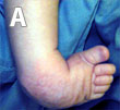
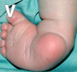
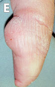
The four components of the deformity which make up a clubfoot can be easily remembered using the acronym ‘CAVE’ which stands for:
A – adduction
V – varus
E – equinus


In approximately 20% of cases clubfoot is non-idiopathic and occurs in association with other neuromuscular deformities such as spina bifida, arthrogryposis and cerebral palsy. Clubfoot has been thought to be associated with developmental dysplasia of the hip (DDH), however, a recent study in the USA found no evidence of an association (28). In any case, it is important that a full physical examination is carried out in children presenting with clubfoot in order to rule out the presence of any other disorders.
Clubfoot must be differentiated from two other disorders: postural equino-varus and metatarsus adductus. Postural equino-varus is caused by the position of the baby in the womb putting pressure on the foot, causing it to be moulded in an abnormal position. On presentation, there may be some degrees of forefoot adduction but it will be flexible and easy to correct. There will be no Achilles tendon contracture. Most cases of postural equino-varus resolve spontaneously but they should be monitored, especially when the child is born prematurely as in these cases a true clubfoot can sometimes present as a postural equino-varus (29).
Metatarsus adductus is a common deformity in childhood in which the forefoot will be adducted and the lateral border of the foot curved. Unlike clubfoot, the heel will not be in equinus and varus. Metatarsus adductus usually resolves spontaneously, but may require casting if the deformity is rigid (29).
The information provided by GCI is based on publications in scientific journals (references indicated by numbers within the text). To see the full reference list click here.


